
UNDERSTANDING PET
What is PET?
Why does calibration matter?
A short introduction to molecular imaging, reference phantoms and the value of a consistent measurement.
01 / MOLECULAR IMAGING
Seeing how
the body functions.
Positron emission tomography (PET) is an imaging method that detects the distribution of a radioactive tracer in the body. Depending on the tracer, it can reveal information about biological activity.
PET is often combined with computed tomography (CT). PET provides information about tracer distribution; CT provides anatomical context.
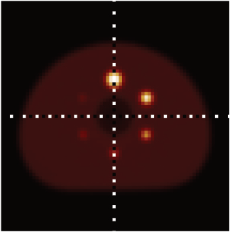 REFERENCE PHANTOM / PET IMAGE
02 / THE NEED FOR CALIBRATION
A common basis
for comparison.
Quantitative PET is used to compare measurements across time, instruments and research sites.
Differences in instrumentation, settings and reference-source geometry can affect the result. A phantom provides a controlled test object with known design and activity concentration, allowing teams to assess performance under defined conditions.
Long-lived solid-state phantoms can be measured repeatedly without preparing a new short-lived liquid source each time. They support quality-control workflows, method development and comparisons across related measurement instruments.
03 / A DIFFERENT APPROACH
From repeated preparation
to a reusable reference.
Conventional fillable phantom
Long-lived solid-state phantom
Preparation and filling of a container with a radioactive liquid increases radiation exposure.
Use a manufactured solid-state body for repeated measurements. There is no need for filling: the radioactive liquid is injected during manufacturing.
Source preparation and change in count rate due to radioactive decay influence workflow.
For long-lived Ge-68 sources, activity stays nearly constant during the comparatively short measurement time, so no significant decay-related change in count rate is expected. Activity is specified at a defined reference date.
The container and non-active interfaces constrain the possible geometry.
Digitally defined geometry enables application-specific and instrumentation-specific structures.
Suitability depends on the isotope, instrument, measurement task and product documentation. Solid-state sources remain radioactive and require appropriate handling.
Questions, answered.
What exactly is a phantom?
A phantom is a test object used to assess an imaging or measurement system. Its controlled geometry and source properties let teams investigate performance without using a patient as the test subject.
Does a phantom replace the radioactive tracer used in a patient?
No. A reference phantom is used to test or evaluate instruments. It has a different purpose from a radiopharmaceutical administered for a patient examination.
Why use Ge-68?
Germanium-68 has a half-life of approximately 271 days and decays to the positron-emitting isotope gallium-68. This makes it useful for long-lived PET reference sources. Product service life also depends on the material, activity and intended use.
What is SPECT?
Single photon emission computed tomography (SPECT) is another nuclear medicine imaging technique. It detects emitted gamma photons to image tracer distribution. SwissNuCRO’s initial product focus is Ge-68 for PET/CT; SPECT sources are a planned development area.
LET’S TALK ABOUT YOUR APPLICATION